Our bimonthly Fundamental Research Update keeps you informed about the latest advances in lab-based ALS research. We break down new studies using cellular and animal models, highlight progress on promising preclinical targets, breakdown trending research news, and showcase Canadian led science. 

For other news on clinical research and trials, read our Clinical Research & Trials Updates. 

ALS Canada is proud to have supported several of the studies below through our Research Program.  

In the lab

*A common feature in most ALS cases is a disruption of the normal function of a protein called TDP-43. This protein is typically found primarily within the nucleus of a cell, where it helps to regulate essential cell processes, but in ALS, it becomes trapped outside in the cytoplasm forming clumps or aggregates. These clumps are theorized to contribute to motor neuron damage and death. Because of this, treatments aimed at preventing or clearing TDP43 clumps could offer a possible way to slow or stop ALS progression. 

Dr. Janice Robertson (University of Toronto) and team have identified a small molecule, called JRMS, that strengthens the activity of a protein called importin-β1 (Kpnβ1) in cells. This protein acts like a cellular chaperone, helping prevent TDP-43* from misfolding and forming clumps, and assisting in its movement back to the nucleus, where it has important functions. In studies using cells and mouse models, JRMS showed that it could prevent new clumps from forming and help dissolve existing ones, while also restoring nuclear TDP-43. 

Dr. Robertson has received follow-up funding through the ALS Canada Discovery Grant program to refine and improve JRMS to make it more druglike, easier to deliver, and able to reach the brain when taken orally. If the molecule continues to show promising results, the team will work to prepare the drug for a path to testing in clinical trials; a process that might take several years. This work could represent an important early step toward creating a therapy that directly targets a very common pathological mechanism in ALS, with the long-term goal of slowing disease progression. 

Additional recent studies also highlight work in TDP-43. This preprint publication by Dr. Alicia Dubinski (Université de Montréal and Dr. Christine Vande Velde’s team, challenges a longstanding hypothesis about the protein. Some researchers believe that TDP-43 mislocalization partly happens because cells are under constant stress and form temporary “stress granules,” which were thought to eventually turn into harmful TDP43 clumps.  

In this supported project, Dr. Vande Velde’s team used mouse models of ALS, exposing them to repeated physical stress (heat) to directly test whether stress granules lead to TDP43 problems. They found that stress granules and TDP43 clumps are separate, reversible structures and do not transform into one another. Surprisingly, even when stress granules were impaired, stress still caused TDP43 to leave the nucleus and build up in the cytoplasm, where it shouldn’t be, eventually leading to the loss of motor neurons. These results suggest that TDP43 damage can happen independently of stress granules, suggesting that therapies targeting cellular stress responses alone may not address the root cause of TDP43 pathology in ALS and related diseases. 


Another study by Dr. Kevin Talbot (University of Oxford) and team closely examined motor neurons derived from mouse stem cells expressing a variant form of the TARDPB (TDP-43) gene at normal levels.  They found that these neurons have trouble moving important materials along their nerve fibres (axonal transport), and produce less energy under normal conditions, surviving less.  

The important finding is that these problems occurred before TDP-43 built up in clumps or mislocalized to the cytoplasm. This suggests that damage to motor neurons may start earlier, due to subtle disruption of TDP-43’s normal role, rather than only after large protein aggregates form. Overall, this study strengthens the overall evidence that loss of normal function is closely linked to TDP-43 dysfunction in ALS and reinforces more recent work that demonstrates impaired function before being mislocalized from the nucleus.  

These findings may carry big implications when developing new treatments targeting TDP-43 biology. 

*Axons are the long “tails” of motor neurons that carry messages inside the brain, and all the way down to the muscles. To keep these neurons healthy, axons need to be able to produce some of their own proteins locally, rather than relying entirely on protein production in the cell body. In ALS, this local protein production in axons may become disrupted, and this breakdown could contribute to disease progression, although researchers are still working to understand exactly how.  

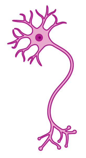
In this study, researchers from Belgium used a powerful lab tool called compartment-specific spatial transcriptomics to map exactly which genes are active in different parts of motor neurons in adult mice, comparing the cell body to the axon*. Specifically, they looked at neurons carrying a pathogenic variant in a known ALS gene called FUS**. They confirmed that FUS-variants disturb the normal set of RNAs found in axons and disrupt the local protein making processOne key protein affected is EIF5A, which is essential for efficient protein production and becomes impaired in neurons making the FUS variant due to a reduction in its active form. 

The researchers then tested a compound called spermidine, which can restore the active form of EIF5A. When they applied spermidine specifically to axons, it improved protein production and reduced the harmful effects caused by FUS variants. They also tested spermidine in fruit fly models of ALS caused by variants in the FUS and TARDPB (TDP43), genes, showing reduced ALS-related toxicity there as well. 

These results suggest that problems with local protein production in axons contribute to ALS, and that boosting this process, possibly with something like spermidine, could be a direction for future therapies. 

**Pathogenic variants (mutations) in the FUS gene are a known risk factor for ALS and contribute to about 1.2 to 1.6% of all ALS cases.

In the gene

*ALS Canada is a proud supporter and has contributed close to 1,000 Canadian samples to Project MinE.  

In this recent study by a team in the Netherlands, using Project MinE* data, researchers combined and carefully standardized genetic data from 22 different research cohorts, creating the largest ALS genetic dataset ever assembled, with more than 17,000 people living with ALS. By analyzing these data and focusing especially on ultrarare genetic variants, the researchers identified new strong candidate genes and gene pathways linked to ALS risk, including, YKT6HTR3C, GBGT1, and KNTC1. They also provide strong, independent validation for genes with limited previous evidence: ARPP21, DNAJC7, and CFAP410. Interestingly, the findings also support the oligogenic idea that ALS risk can build up from multiple small genetic variants, a concept we’ve highlighted in a previous Research Update. 

The results also indicate that a genetic contribution can be identified in approximately 25% of people with ALS, regardless of a family history, where previous data estimated about 20% of cases. These findings expand our understanding of how rare genetic changes contribute to ALS and provide new leads for future research and potential therapies. 

This review article by Dr. Matti Allen (University of Ottawa) and others, including the ALS National Genetic Counsellor Maya Binet, highlights a lesser-known group of ALS cases caused by autosomal recessive genetic variants, which are often linked to earlier onset or atypical symptoms. By detailing key genes and biological pathways involved, the authors show how understanding these forms of ALS could improve diagnosis, guide prognosis, and support the development of future gene-targeted therapies. 

In the papers

This recent article talked about findings by Dr. Aaron Burberry (Case Western Reserve University) and team. In their study, the researchers found that some gut bacteria prevalent in C9orf72-ALS* make special forms of glycogen, a stored sugar, that immune cells can recognize as inflammatory. 

In mice with variants in the C9orf72 gene, the authors found that these bacterial glycogens accumulate in the gut, amplifying immune responses, weakening the blood–brain barrier, and promoting immune cell entry into the brain and spinal cord. Breaking down this glycogen in the gut reduced brain inflammation and improved survival in these mouse models.  

In humans, the authors analyzed stool samples, finding this inflammatory glycogen in 68% people living with C9-ALS and one person with C9-FTD, although it also appeared in over 30% of healthy individuals.  

The bottom line: the authors showed that glycogen triggers an exaggerated immune response, but only in mice lacking normal C9orf72 function. In humans, they only showed that the same type of glycogen exists in the gut. Therefore, more research is still needed before we can draw firm conclusions about how glycogen and the gut–brain connection influence ALS and FTD. However, this remains an important area of study, and this project adds valuable insight to our growing understanding of the gut-brain connection in ALS. 

*Pathogenic variants (mutations) in the C9orf72 gene are a known risk factor for ALS and contribute to about 10% of all ALS cases. 

Congratulations to Dr. Alicia Dubinski on receiving the prestigious Schmidt Science Fellowship, a welldeserved recognition! We were proud to support Dr. Dubinski earlier in her career through an ALS Canada Trainee Award, where she worked on TDP43 pathology and stress granules, key areas of ALS research. We’re excited to see her continue this important work in the U.S., contributing to progress across ALS and other neurodegenerative diseases. 

In a glance

Oral microbiome differences offer clues to ALS subtypes  

In this supported study, researchers led by Dr. Gerald Pfeffer (University of Calgary) analyzed saliva samples from people living with ALS and healthy controls, recruited from Canada and South Korea, and found that different ALS subtypes showed distinct patterns of oral bacteria. Bifidobacterium was more common in people with bulbar-onset ALS, while Haemophilus was more common in those whose symptoms began in the limbs. While no major differences were found between people with ALS and healthy controls overall, noticeable differences appeared between participants from different countries and even between households, suggesting that environmental, lifestyle, and genetic factors may all play a role. However, it is important to note that oral health–related factors (such as clearance of secretions and hygiene) are difficult to fully control and may influence results, underscoring the need for further studies. 


Compensatory scaling of modulatory neural populations in response to motor challenges 

This supported study by Dr. Turgay Akay’s team (Dalhousie University) shows how different brain and spinal cord signaling systems work together to fine-tune movement, adjusting muscle activity based on speed and demand. The researchers also found that in ALS mouse models, these systems are recruited differently over time, suggesting the nervous system tries to compensate as motor neurons become impaired, offering insight into how complex motor control changes during disease progression. 

Age and life stage in the experience of amyotrophic lateral sclerosis: a scoping review.  

This review article by Dr. Andrea Parks (University of Toronto) looked at how age and life stage shape the experiences of people living with ALS, analyzing more than 40 studies published over the past decade. While many aspects of living with ALS differ by age, the authors found that important factors such as life transitions and age-related roles (like work or caregiving) are often overlooked, highlighting a need for more personalized, age-aware approaches to ALS care. 


Data-driven disease subgrouping in ALS: a multicenter cerebral functional connectivity study 

This study by Dr. Sanjay Kalra’s team (University of Alberta), using CALSNIC data, found that brain imaging can objectively detect differences in brain connectivity among people living with ALS that align with disease stage and real-world functioning. Using data-driven imaging approaches identified clearer and more widespread brain changes than traditional clinical grouping methods, suggesting neuroimaging could serve as a valuable biomarker for patient stratification and clinical trial design. 

Support the cause

You can make a difference.

Donations provide crucial services and information to people living with ALS and allow us to invest in research and advocacy dedicated to improving quality of life.